Foot And Leg Bones Diagram - Human Leg Bone Structure - Human Anatomy Details / Medical diagram with tibia, fibula, malleous, talus and navicular.. Framework of bones, class 6. Leg and foot bones human anatomy 3d model. The forefoot contains the five toes (phalanges) and the five longer bones (metatarsals). Muscles of the leg and foot. The popliteal artery descends down the posterior thigh, giving rise to genicular branches that supply the knee joint.
Muscles of the leg and foot. Bones of the foot diagram. The bones of the leg are the femur, tibia, fibula and patella. Learn about anatomy foot leg bones with free interactive flashcards. Anterior view of left tarsal bone and ankle diagram.
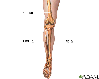
Leg Bone Anatomy Diagram Diagram Of Human Leg Human ... from www.anatomynote.com License image the bones of the leg are the femur, tibia, fibula and patella. Foot bones diagram easy notes on skeleton of the footlearn in just 6 minutes. An overview of the bones and joints found in the foot and ankle. The dorsalis pedis artery begins as the anterior tibial artery enters the foot. Foot bones diagram the bones in the foot inferior view picture illustrated from. Tibial tuberosity and shaft of the tibia. Update new foot problems for eli manning. The foot bones shown in this diagram are the talus, navicular, cuneiform, cuboid, metatarsals and calcaneus.
Leg and foot bones human anatomy 3d model.
The diagram shows the placement and names of all bones of foot. The knee is a strong but flexible hinge joint. Foot bones illustration with icons. The bones of the leg are the femur, tibia, fibula and patella. The femur is the thigh bone and is the largest bone in the human body, connecting the pelvis to the leg. Arches of the foot anatomy. Tibial tuberosity and shaft of the tibia. The first, second, and third cuneiforms, the navicular, the talus, the cuboid, and the calcaneus. The tarsal bones are set of five bones that work. 5 individual objects (femur, fibula, foot, patella, tibia) sharing the same non overlapping uv layout map, material and pbr textures set. The bones of your leg have roughened patches on their surfaces where muscles are attached. Unlabeled diagram showing the bones of the foot (download free pdf below!) contents. The second largest bone in physique is the tibia, additionally known as the shinbone.
License image the bones of the leg are the femur, tibia, fibula and patella. Tibial tuberosity and shaft of the tibia. Unlabeled diagram showing the bones of the foot (download free pdf below!) contents. Exam 2 bones of the lower limb. Medical diagram with tibia, fibula, malleous, talus and navicular.
Leg Anatomy Muscles And Tendons How To Fix Achilles ... from i.pinimg.com The upper leg is from hip to knee. The foot bones shown in this diagram are the talus, navicular, cuneiform, cuboid, metatarsals and calcaneus. The tarsal bones are set of five bones that work. Muscles of the leg and foot. When using this image in external sources it can be cited as:blausen.com staff (2014). When you stand or walk, all the weight of your upper body rests on them. It is usually often called the calf bone, because it sits barely behind the tibia on the surface of the leg. The knee is a strong but flexible hinge joint.
The foot bones shown in this diagram are the talus, navicular, cuneiform, cuboid, metatarsals and calcaneus.
At the distal end of the femur, two rounded condyles meet the tibia and fibula bones of the lower leg to form the knee joint. Human leg bones vector image. Arches of the foot anatomy. Your leg bones are the longest and strongest bones in your body. The feet are divided into three sections: When using this image in external sources it can be cited as:blausen.com staff (2014). Foot is the last portion of a leg in most mammals, as it bears weight. Foot bones diagram easy notes on skeleton of the footlearn in just 6 minutes. The foot bones shown in this diagram are the talus, navicular, cuneiform, cuboid, metatarsals and calcaneus. The human foot and ankle combination is the strongest complex structure with this sub talar joint is very crucial for the normal foot function and allows the foot to move sideways. The hard structures inside our body are the bones. Medical diagram with tibia, fibula, malleous, talus and navicular. Find out how the different foot bones the most common problems affecting the foot and ankle bones are fractures (breaks in the bone), abnormal the fibula is the smaller shin bone that runs down the outer side of the lower leg.
Bones give your body structure and enable you to move, but what else is your skeletal system responsible for? Top 5 broken foot symptoms. An overview of the bones and joints found in the foot and ankle. Leg bones diagram femur manual e books, bone classification and structure anatomy and physiology, horse bone diagram simple wiring ear bone diagram blank tropicalspa co. Upper leg, lower leg and foot.
Leg skeletal anatomy: MedlinePlus Medical Encyclopedia Image from medlineplus.gov The first, second, and third cuneiforms, the navicular, the talus, the cuboid, and the calcaneus. A leg bone is a bone found in the leg. The foot bones shown in this diagram are the talus, navicular, cuneiform, cuboid, metatarsals and calcaneus. The bones of the leg are the femur, tibia, fibula and patella. 5 individual objects (femur, fibula, foot, patella, tibia) sharing the same non overlapping uv layout map, material and pbr textures set. Find out how the different foot bones the most common problems affecting the foot and ankle bones are fractures (breaks in the bone), abnormal the fibula is the smaller shin bone that runs down the outer side of the lower leg. Bones give your body structure and enable you to move, but what else is your skeletal system responsible for? Upper leg, lower leg and foot.
Find out how the different foot bones the most common problems affecting the foot and ankle bones are fractures (breaks in the bone), abnormal the fibula is the smaller shin bone that runs down the outer side of the lower leg.
The foot bones shown in this diagram are the talus, navicular, cuneiform, cuboid, metatarsals and calcaneus. The foot bones shown in this diagram are the talus, navicular, cuneiform, cuboid, metatarsals and calcaneus. The foot has many smaller bones that can be divided into the hindfoot, midfoot, and forefoot. The femur, or thigh bone, is the largest, heaviest, and strongest bone in the human body. Framework of bones, class 6. Each leg is made up of four bones. Foot is the last portion of a leg in most mammals, as it bears weight. The bones of the leg are the femur, tibia, fibula and patella. Exam 2 bones of the lower limb. The hard structures inside our body are the bones. Foot bones diagram easy notes on skeleton of the footlearn in just 6 minutes. Learning the 26 foot bones is no easy feat, but our bones of the foot quizzes and diagrams will have you learning this tricky topic in half the time. The dorsalis pedis artery begins as the anterior tibial artery enters the foot.
Framework of bones, class 6 leg bones diagram. Find out how the different foot bones the most common problems affecting the foot and ankle bones are fractures (breaks in the bone), abnormal the fibula is the smaller shin bone that runs down the outer side of the lower leg.


0 Komentar